Elbow Anatomy
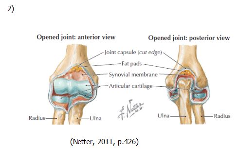
Type of joint: the elbow is made up of three joints. The humeroulnar articulation (the synovial hinge joint with articulation between the trochlea of the humeral condyle and the trochlear notch of the ulna) and the humeroradial articulation (the articulation between the capitulum of the humeral condyle and the concavity on the superior aspect of the head of the radius). The third is a pivot-type synovial joint with articulation between the head of the radius and the radial notch of the ulna (Medscape, 2013) (fig. 1 and 2).
Radial head: the rounded proximal end of the radius. It articulates with the ulna (Gest & Schlesinger, n.d.) (fig.5).
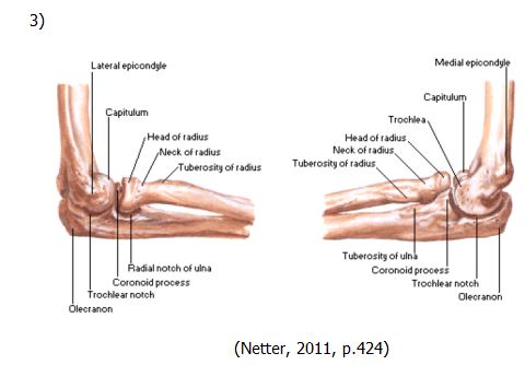
Radial neck: constricted area of the radius located distal to the head (Gest & Schlesinger, n.d.) (fig.3).
Radial tuberosity: insertion site for the biceps brachii. The radius is located distally to the neck (Gest & Schlesinger, n.d.) (fig.3 and 5).
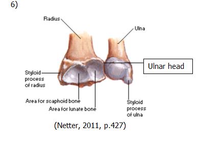
Styloid process of radius: projects laterally to the proximal row of the carpals (Gest & Schlesinger, n.d.) (fig.6).
Ulna styloid process: projects from the distal surface of the head of the ulna. The articular disk and the distal radioulner joint attach here (Gest & Schlesinger, n.d.) (fig.6).
Ulnar head: located at the distal end. It articulates with the radius (Gest & Schlesinger, n.d.) (fig.6).
Ulnar tuberosity: gives insertion to part of the brachialis (Gest & Schlesinger, n.d.) (fig.3).
Coronoid process: anterior projection located distally to the trochlear notch and is the site of insertion for the brachilais muscle (Gest & Schlesinger, n.d.) (fig.3).
Olecranon: proximal end of the ulna and is the insertion site for the triceps brachii tendon. With the proximal portion of the coronoid process, it forms a depression that articulates with trochlea. This articulation allows for anteroposterior plane motion (fig.3) (Gest & Schlesinger, n.d.).
Synovial membrane: membrane in the elbow that produces synovial fluid (Thomas, L. (September 9, 2013). Lecture 1: Orientation to Musculoskeletal Ultrasound [PowerPoint]). It allows movement of the elbow joint (Gest & Schlesinger, n.d.) (fig.2).
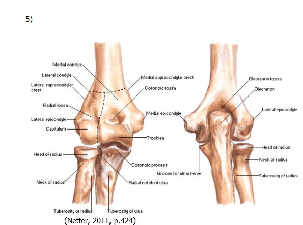
Humerus trochlea: located at the medial distal end of the humerus. It articulates with the trochlear notch of the ulna (Gest & Schlesinger, n.d.) (fig. 5).
Capitulum: part of the humerus that caps the distal end of the lateral condyle. It articulates with the radius’s head (Gest & Schlesinger, n.d.) (fig.3 and 5).
Medial epicondyle: protects the ulnar nerve. It is a rounded projection at the distal and medial end of the humerus. Muscles that flex the forearm and wrist attach here (Body Smart, n.d.) (fig.1 and 3).
Lateral epicondyle: a rounded projection at the distal and medial end of the humerus. Muscles that flex the forearm and wrist attach here (Body Smart, n.d.) (fig.1 and 3).
Medial supracondylar ridge: runs from the medial epicondyle of the humerus proximally (Gest & Schlesinger, n.d.) (fig.5).
Lateral supracondylar ridge: runs from the lateral epicondyle of the humerus laterally (Gest & Schlesinger, n.d.) (fig. 5).
Coronoid fossa: a depression on the anterior surface of the humerus; it is located proximally to the trochlea near the elbow and accommodates the coronoid process of the ulna when one flexes their elbow (Gest & Schlesinger, n.d.) (fig.5).
Radial fossa: a depression on the anterior surface of the humerus. It is located proximally to the capitulum. It accommodates the radial head when one flexes their elbow (Gest & Schlesinger, n.d.) (fig.5).
Olecranon fossa: depression on the posterior surface of the humerus and is located proximally to the elbow. It accommodates the olecranon process of the ulna when the elbow is flexed (Gest & Schlesinger, n.d.)(fig.5).
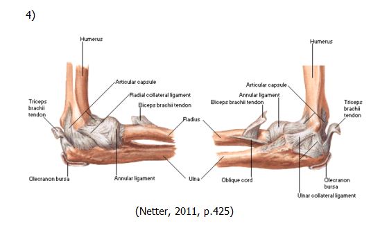
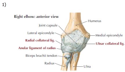
Annular ligament: surrounds the head of the radius and keeps it in the radial notch of the ulna. It attaches the anterior margin of the radial notch of the ulna and goes around the radial head to insert posteriorly on the notch (Gest & Schlesinger, n.d.) (fig.1).
Lateral ulnar collateral ligament: joins the distal aspect of the humerus to the proximal part of the ulna (Gest & Schlesinger, n.d.) (fig.1).
Radial collateral ligament: spans the lateral side of the elbow joint. Connects the lateral epicondyle of the humerus to the radius and the annular ligament. Its function is to reinforce the lateral side of the articular capsule of the elbow (Gest & Schlesinger, n.d.) (fig.1).
Tendon of the biceps brachii: inserts on the radial tuberosity (Jacobson, 2013, p. 72). (fig.1 and 4).
Brachialis: originates on the anterior distal half of the humerus and inserts on the coronoid process and tuberosity of the ulna. It flexes the forearm at the elbow (Body Smart, n.d.).
Olecranon bursa: found between the point of the elbow and the skin. It allows for movement (Gest & Schlesinger, n.d.) (fig.4).
Common extensor lateral: located on the lateral epicondyle of the humerus. It is the attachment site for the extensor carpi radialis brevis, the extensor digitorum, the extensor digiti minimi and the extensor carpi ulnaris (Anatomy TV, n.d.).
Common felxor origin medial: originates at the medial epicondyle of the humerus. Allows for flexion of the phalanges, metacarpophalangeal joints and wrist (Anatomy TV, n.d.).
Nerves-
Ulnar nerve: runs in the space between the olecranon process of the ulna and the medial epicondyle (Jacobson, 2013, p. 73).
Median nerve: found between the ulner and humeral heads of the pronator teres (Jacobson, 2013, p. 73).
Interosseuous nerve: begins anterior to the lateral epicondyle of the humerus. From there, it enters the cubital fossa and goes into the supinator to reach the forearm's extensor compartment (Anatomy TV, n.d.).
(Netter, 2011, p.426)